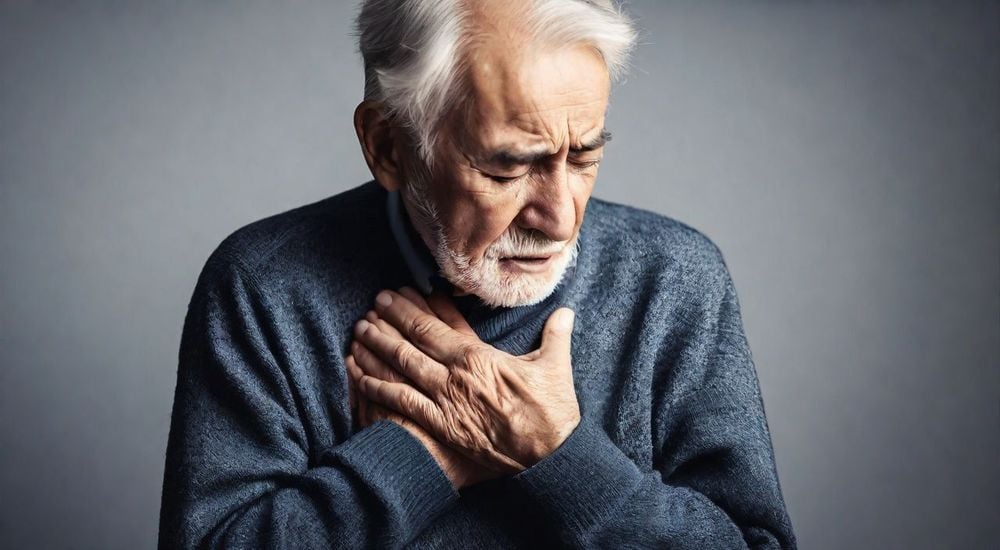
Arrhythmia is an abnormal heart rhythm that can occur at any age and at any time. Symptoms of arrhythmia include palpitations, chest pain, a feeling of loss in the chest, chest pain, or shortness of breath. The following article will help people better understand and have a plan to prevent and stop arrhythmia from progressing to a more serious condition.
1. Overview of arrhythmia
1.1 How is heart rhythm formed?
Normally, the human heart has a total of 4 chambers: 2 smaller chambers located at the top called the atria, and 2 larger chambers located at the bottom called the ventricles.
A normal heart rhythm is created by a structure located in the heart, called the sinoatrial node, located in the right atrium. The electrical impulses generated by the sinoatrial node will spread to the atria of the heart, then this impulse will be transmitted to the ventricles through the atrioventricular node and the conduction bundles.
These electrical impulses are emitted by the SA node and transmitted throughout the heart continuously, creating the heartbeat. The formation and propagation of these electrical impulses play an important role in controlling the heartbeat and creating the heart rhythm.
A normal heart rhythm is created by the SA node, so it is also called sinus rhythm. The frequency of the sinus rhythm (the number of heartbeats per minute) is not fixed, but will change based on the physiological state, body activity and environmental conditions.
1.2 What is heart rate?
Heart rate, also known as heart rate, is the number of times the heart beats in one minute. The heart rate of a normal person ranges from 60 to 100 times per minute. However, heart rate can vary greatly because it is affected by many different factors. For example, after a big meal, exercise, fever, or in emotional states such as anger, fear, or excitement, the heart rate may increase more than normal (>100 beats/minute). Conversely, the heart rate may be slower than normal when you are sleeping or in people who have exercised (maybe lower. These variations are called physiological, because they depend on activity level, mood, overall health, and environmental conditions.
You can check your heart rate yourself using 3 methods:
● Pulse counting: Place your finger on your wrist (near your thumb), on the inside of your forearm, or on your neck (angle of your jaw) and count the number of times your heart beats in 1 minute.
● Listen with a stethoscope: Use a stethoscope under the guidance of a medical professional to listen to your heartbeat.
● Use an electronic device: Devices such as watches, smartphones, blood oxygen saturation monitors, or blood pressure monitors with built-in heart rate measurement functions make it easy to check your heart rate level.
You can choose which testing method is best for you, depending on your habits personal and most convenient for you.
1.3 What is a normal heart rate?
A normal heart rate is a heartbeat that originates from the sinus node (Sinoatrial) and is conducted to cause atrial depolarization, then transmitted through the AV node/His-Purkinje system to cause ventricular depolarization to help the ventricles. This activity occurs rhythmically, forming cycles and is relatively regular.
When sinus rhythm is normal:
● The resting heart rate will fluctuate between 60 – 100 cycles / minute.
● Normal sinus rhythm does not cause any symptoms, the heart beats fast and feels nervous in some cases: increased emotions, anxiety, fever, … and these symptoms will return to normal quickly.
1.4 What is arrhythmia?
● Arrhythmia is an abnormal electrical condition of the heart, including abnormalities in the process of rhythm generation and electrical conduction in the heart chambers. Arrhythmia includes the following clinical manifestations: heart rate is too fast (frequency > 100 beats/minute) or too slow (frequency < 60 beats/minute), irregular heart rate, sometimes the heart beats fast, sometimes the heart beats slow.

Heart rate that is too fast or too slow are clinical manifestations of arrhythmia.
● In some cases, arrhythmia does not show symptoms or only appears as a feeling of nervousness, a feeling of palpitations, a fast or irregular heartbeat. However, in many cases, arrhythmia can be life-threatening, requiring the patient to be hospitalized for emergency care.
● Arrhythmia is a common disease and often occurs in daily practice. The disease can be detected when the patient goes for a general health check-up or when they visit other specialists. A large number of elderly patients when hospitalized for treatment of diseases such as diabetes, high blood pressure… have discovered that they have arrhythmia, and there are even cases where atrial fibrillation is detected when the patient is hospitalized due to a stroke.
2. Causes of arrhythmia
Arrhythmias can have many different causes. They can result from abnormalities and diseases of the heart, or they can arise from problems in other organs that affect the heart rhythm (for example, thyroid disease, kidney failure causing electrolyte disturbances).
Arrhythmias can appear briefly, lasting only a few minutes or less, or they can appear in episodes without warning. However, some types of arrhythmias can last for hours, or even years.
Causes of arrhythmias include:
● Abnormal activity and weakness of the sinus node.
● The presence of another abnormal rhythm source in the heart.
● The presence of abnormal electrical pathways in the heart.
● Blockage of the heart’s conduction system.
● Damage to the heart muscle.
● Electrolyte disturbances in the body.
● Effects of other medications or toxic compounds.
● Effects of abnormalities in other organs on the heart (for example, thyroid problems).
3. Symptoms of arrhythmia
Arrhythmia in some patients does not cause unpleasant symptoms, especially in cases of chronic arrhythmia, in which the patient may not recognize the symptoms of the disease.
However, in some cases, arrhythmia will cause more serious symptoms, notably including:
- Palpitations: This is a typical and common symptom of arrhythmia. The patient may feel the heart beating strongly in the chest, the patient feels like the heart stops beating for a moment and beats strongly again. Patients often describe this symptom as a feeling of nervousness or palpitations.
- Sudden shortness of breath: This symptom is often accompanied by a feeling of irregular heartbeat or a feeling of nervousness. Shortness of breath is one of the signs suggesting the risk of myocardial infarction or dangerous arrhythmia.
- Dizziness: the patient feels dizzy, everything around is spinning, the person loses balance. Dizziness is often a symptom of many different diseases, including arrhythmia.
- Fainting: the patient suddenly loses consciousness for a short time. This is a worrying symptom, because it can lead to serious injuries if the patient faints while participating in traffic or going up the stairs. Today, there are many modern methods to diagnose and treat dangerous arrhythmias, including tilt table testing and electrophysiological investigation techniques, as well as Radio Frequency (RF) ablation therapy to help diagnose and treat these conditions.
4. Subjects susceptible to arrhythmia
Arrhythmia can occur at any age and gender, however, according to research results, the disease is more likely to occur in the following subjects:
● People over 60 years old.
● Patients with a history of high blood pressure.
● People with coronary artery disease.
● Patients with heart failure.
● Heart valve disease.
● People who have had open heart surgery.
● People with sleep apnea syndrome.
● Thyroid disease.
● Diabetes.
● Chronic lung disease.
● Drinking a lot of alcohol and using stimulants.
● People over 60 years old who have had infections or internal diseases.

People over 60 years old are susceptible to arrhythmia
5. Types of arrhythmias
5.1 Types of arrhythmias
5.1.1 Paroxysmal supraventricular tachycardia
● Paroxysmal supraventricular tachycardia is the most common type of tachycardia and can occur at any age. The origin of the tachycardia can originate from atrial foci, atrioventricular accessory pathways, or from the atrioventricular node.
● The heart rate can occur suddenly, even when the patient is resting or sleeping. The heart rate during a tachycardia episode usually ranges from 150 to 210 beats/minute and is regular. During a tachycardia episode, the patient often experiences mild symptoms such as palpitations, chest discomfort, shortness of breath, or weakness. However, in some cases, more severe symptoms may appear with manifestations such as dizziness, lightheadedness, low blood pressure, and fatigue.
● Usually, paroxysmal supraventricular tachycardia does not have a major impact on the patient’s overall health, because it occurs infrequently and improves on its own within a few hours. However, in some cases, tachycardia can occur more frequently, last longer, cause more symptoms, and in these cases, the patient needs to be treated by a specialist in the field of arrhythmia.
● Treatment for this condition will depend on the type of tachycardia, the severity of symptoms, the frequency of tachycardia, the patient’s overall health, and the patient’s choice between direct investigation and treatment (such as investigation and arrhythmia ablation) or the use of drugs to control and reduce tachycardia.
5.1.2 Atrial flutter
● Atrial flutter is a type of tachycardia that occurs in the atria of the heart, usually formed by one or more electrical conduction circuits under the atrial structure. In this case, the atria contract rapidly and regularly at a frequency ranging from 240 to 340 times/minute.
● Although the atria contract rapidly, the impulse during conduction through the AV node is reduced before being transmitted to the two ventricles below. This is a physiological property of the AV node to protect the ventricles from the effects of arrhythmias in the atria.
● Symptoms of atrial flutter are similar to those of other supraventricular tachycardias, including palpitations, chest pain, shortness of breath, weakness, and dizziness.
● Atrial flutter rarely causes fainting or fainting. However, in some cases, the patient may experience stroke symptoms, such as weakness, paralysis of the arms/legs, possible slurred speech, or loss of consciousness due to blockage by a blood clot. Patients after a stroke are often diagnosed with atrial flutter or atrial fibrillation.
● Some factors that increase the risk of atrial flutter include: old age, obesity, alcoholism, heart valve disease, congenital heart disease, or patients who have had heart surgery.
5.1.3 Atrial Fibrillation
● Atrial fibrillation is one of the most complex types of arrhythmias in the atrial layer of the heart. In atrial fibrillation, the atria operate out of sync in many different areas, creating rapid, irregular, and chaotic impulses throughout the atria. The result of these abnormalities is a rapid and irregular heart rhythm.
● Atrial fibrillation can occur transiently in episodes without causing any obvious symptoms. However, as the disease progresses, atrial fibrillation can become continuous, becoming a persistent condition and causing a decline in heart function over time. The symptoms of atrial fibrillation are similar to those of atrial flutter.
● There are many factors and causes that contribute to the increased risk of atrial fibrillation, including: high blood pressure, coronary artery disease, heart valve disease, congenital heart disease, thyroid disease, metabolic disorders (such as diabetes), sick sinus syndrome, chronic obstructive pulmonary disease, sleep apnea syndrome, after heart surgery, older age and overweight obesity.
● Similar to atrial flutter, atrial fibrillation also has the risk of creating blood clots in the heart, causing stroke due to blood clots blocking cerebral vessels. Therefore, doctors need to assess this risk early and perform anticoagulant treatment (if necessary) to prevent stroke. This is extremely important in addition to treating and controlling atrial fibrillation and heart rate in atrial fibrillation.
● Treatment of atrial fibrillation will involve controlling the heart rate and heart rate. In rhythm control, doctors will try to maintain a normal rhythm (sinus rhythm) for patients by using drugs, interventional survey and atrial fibrillation ablation (if necessary), to minimize the recurrence of atrial fibrillation. In heart rate control, doctors focus on keeping the ventricular heart rate from being too fast, regardless of when atrial fibrillation recurs.
● The choice of treatment for atrial fibrillation often depends on many factors and the specific condition of each patient, there is no general treatment method for all. Therefore, when atrial fibrillation is detected, patients need to consult a doctor specializing in arrhythmias to have appropriate treatment options and reduce the risk of the disease getting worse.
5.1.4 Ventricular tachycardia
● Ventricular tachycardia is an arrhythmia originating from the ventricles of the heart. Patients need to pay special attention as soon as ventricular tachycardia appears for the first time. Because ventricular tachycardia has the potential to cause significant harm to health compared to normal ventricular tachycardia and has the risk of serious conditions.
● Moreover, ventricular tachycardia is also a sign of other dangerous cardiovascular diseases that the patient has not yet diagnosed. Given the complexity and danger of ventricular tachycardia, when this condition is discovered, the patient must seek advice from specialists immediately
● Ventricular tachycardia may have no obvious symptoms and only appear transiently, or the patient may only feel lightheaded, have palpitations, or feel unwell. However, as the condition worsens, most patients will experience mild to severe symptoms, including palpitations, dizziness, severe chest pain, shortness of breath, seeing unusual lights, low blood pressure, near-unconsciousness, and fainting.
● Ventricular tachycardia can originate from many causes, including coronary artery disease, heart muscle disease (hypertrophic cardiomyopathy or dilated cardiomyopathy), inherited arrhythmias (long QT syndrome, catecholamine-associated ventricular tachycardia), electrolyte disturbances, side effects of treatment drugs, use of addictive substances such as cocaine or methamphetamine, and finally ventricular tachycardia of unknown cause.
5.1.5 Ventricular fibrillation
● Ventricular fibrillation is the most dangerous form of tachyarrhythmia, directly and immediately threatening the patient’s life, requiring immediate emergency treatment.
● When a patient has ventricular fibrillation, the ventricles of the heart contract very quickly, chaotically, and completely out of sync, causing the heart’s ability to pump blood and maintain circulation to be interrupted. Therefore, when suffering from ventricular fibrillation, the patient has a high mortality rate if not treated promptly, and in case of delayed treatment, there is a possibility of permanent brain damage.
● Symptoms of ventricular fibrillation progress very quickly over time from the time the disease begins. In the first few seconds when ventricular fibrillation begins, the patient often experiences dizziness, sees unusual light, dark eyes, and feels like the limbs are losing strength. If the ventricular fibrillation lasts for nearly 10 seconds, the patient may lose consciousness briefly. When the ventricular fibrillation lasts longer, the patient may faint and organs sensitive to anemia (such as the brain…) begin to be damaged. In this case, if the patient is not treated promptly and ventricular fibrillation persists, the mortality rate is very high.
● There are a number of risk factors that can contribute to ventricular fibrillation, including: acute myocardial infarction, cardiomyopathy, severe inherited ventricular arrhythmias (Brugada syndrome, long QT syndrome), acute myocarditis or injury, severe electrolyte disturbances, overdose of specific drugs such as cocaine and methamphetamine, and ventricular fibrillation of unknown cause.
● Treatment of ventricular fibrillation is a medical emergency and requires immediate treatment. In addition, it is necessary to quickly find and correct the causative factors to help stabilize the patient and prevent recurrence of ventricular fibrillation. Except in cases of ventricular fibrillation due to acute causes, it can be treated to completely recover.
● To prevent sudden death due to recurrent ventricular fibrillation, the only treatment is to place a defibrillator to shorten the ventricular fibrillation and protect the patient’s life. This is not a treatment to eliminate ventricular fibrillation, but an emergency measure to prevent sudden death.
5.2 Bradyarrhythmias
5.2.1 Sinus node failure
● A normal heart rate is one that originates from the sinus node. When the sinus node is malfunctioning, it can lead to a decrease in the ability to generate a steady heartbeat, resulting in bradycardia. This often occurs when the sinus node is poorly adapted to physiological changes or daily activities. For example, the heart rate may not increase or remain slow when the patient is active. In some cases, after the patient experiences a rapid arrhythmia (such as atrial fibrillation), the heart rate becomes very slow or may stop for a long time (about 5 seconds).
● Sinus node failure often progresses slowly, possibly over many years, so the patient may adapt to a slow heart rate without noticing obvious symptoms.
● However, in the presence of symptoms, common signs in patients with SA node failure include: palpitations, feeling that the heart is beating slowly and strongly, frequent fatigue, dizziness, shortness of breath, near-fainting or even fainting.
● Causes or risk factors that contribute to SA node failure or SA node inhibition may include: advanced age, coronary artery or heart muscle disease, inflammatory or infectious diseases affecting the heart, SA node inhibition due to the use of cardiovascular medications (such as blood pressure medications, heart failure medications, coronary artery disease medications, tachyarrhythmia medications), damage to the SA node due to heart surgery, and even some medications used to treat Alzheimer’s disease.
● Treatment of SA node failure depends primarily on whether the patient has mild or severe symptoms, the patient’s limited mobility, and the results of an electrocardiogram (ECG) that shows prolonged sinus pauses. When the sinus node has deteriorated and the heart rate has slowed significantly, medication is rarely effective. In this case, placing a permanent pacemaker is the only way to maintain a normal heart rhythm and prevent sudden death due to prolonged cardiac arrest.
5.2.2 Atrioventricular conduction block
● To maintain a normal heart rhythm, the impulse generated from the sinus node must be transmitted through the conduction pathways from the atria to the ventricles (called atrioventricular conduction) to ensure that this impulse is completely transmitted from the atria to the ventricles. However, if the atrioventricular conduction pathway is damaged at important locations, this can lead to conduction block. In this condition, the impulse cannot be transmitted to the ventricles completely, and if it is completely blocked, it will cause cardiac arrest.
● Depending on the degree of atrioventricular conduction obstruction, the doctor will determine different clinical levels, from mild (grade 1) to severe (grade 3). Patients often experience symptoms similar to other bradyarrhythmias.
● Causes and risk factors contributing to atrioventricular conduction block may include: acute myocardial infarction and coronary artery disease, cardiomyopathy, congenital heart disease, post-cardiac surgery, after percutaneous cardiac intervention procedures that may cause damage to the conduction system, conduction pathway degeneration, cardiotoxic chemotherapy, and many other causes.
● Currently, treatment of atrioventricular block mainly focuses on finding and treating the underlying causes that may lead to the disease. In cases of atrioventricular block without underlying causes, there are almost no drugs to improve the disease. When a patient has severe AV block, high-grade AV block, or symptoms, the last resort is to place a permanent pacemaker to maintain a stable heart rhythm.
5.3 Other disorders
Here are some types of arrhythmias that originate from isolated arrhythmias in the atria or ventricles, occurring alternately with normal sinus rhythm:
● Atrial extrasystoles;
● Ventricular extrasystoles.
Patients with extrasystoles often experience the following symptoms:
● Palpitations;
● Feeling of irregular heartbeat, skipping beats from time to time;
● Feeling of shortness of breath or gasping for air;
● Chest discomfort;
● Shortness of breath;
● There may be limited mobility in some cases.
The doctor may recommend that the patient monitor the Holter ECG within 24 hours to determine the frequency and severity of extrasystoles. Depending on the severity and symptoms of the patient, the doctor will choose the appropriate treatment method. Currently, premature ventricular contractions can be treated through cardiac catheterization and arrhythmia ablation. Each treatment method has its own advantages and disadvantages, and the doctor will plan the appropriate treatment based on the patient’s specific situation.

People with arrhythmia often feel nervous.
6. Complications of arrhythmia
Some mild arrhythmias usually do not significantly affect health. However, other arrhythmias, especially those that cause symptoms as described earlier, can cause serious consequences, including:
● Stroke: People with arrhythmia have a 5 times higher risk of stroke than healthy people. This is because when you have an arrhythmia, blood does not circulate strongly enough to the upper body, increasing the risk of blood clots forming. These clots can travel to the brain, block blood vessels and cause a stroke.
● Limitation of mobility and daily activities: In order to perform daily activities, the body needs a constant supply of oxygen-rich blood. However, when you have an arrhythmia, your heart cannot pump enough blood to other parts of your body, causing constant fatigue and weakness.
● Heart failure: The heart is responsible for pumping blood to other parts of your body to keep you alive. However, an arrhythmia can prevent your heart from pumping blood to where it needs to go effectively. This makes your heart work harder and gradually weakens it. This prevents your heart from functioning properly and leads to heart failure.

Arrhythmias can lead to heart failure.
● Sudden death: Some forms of arrhythmia can cause hidden danger, with no symptoms or transient symptoms that are not obvious. However, when a patient experiences a severe arrhythmia, it can cause sudden death. One of the main causes of sudden death in young people is severe arrhythmia, which is likely caused by a genetic mutation.
7. Methods of diagnosing arrhythmia
To diagnose arrhythmia, an arrhythmia specialist will conduct a series of tests and evaluate your cardiovascular condition.
Diagnostic methods may include: a general cardiovascular examination, information about your medical history and the symptoms you are experiencing. The doctor will conduct the necessary tests to determine and assess the severity of the arrhythmia (if any). After conducting a clinical examination and evaluating the test results, your doctor will advise you on your condition and develop a suitable treatment plan.
It is important to note that some arrhythmias may appear intermittently or only transiently. This means that when you visit your doctor, if at that time your heart is in a normal state (because the arrhythmia has improved on its own), the clinical examination will have normal results and will not detect any abnormal signs. Therefore, if you suspect an arrhythmia based on your symptoms, you should see an arrhythmia specialist as soon as possible, especially if you are experiencing symptoms.
Clinical tests commonly used to diagnose an arrhythmia include:
● Electrocardiogram (ECG): During an ECG, electrical sensors (electrodes) are placed on your chest, arms or legs to record the electrical activity of your heart. This method measures the timing of each electrical phase of the heartbeat.
● 24-hour Holter ECG: Can be worn for one or more days to record the heart’s activity over a longer period of time, increasing the chance of detecting an arrhythmia. Holter ECGs are useful for assessing symptoms associated with arrhythmias as well as monitoring your response to treatment. They are also used to screen and identify people at risk for arrhythmias so they can be treated promptly.
● Event recorder: Used to detect sporadic arrhythmias. The device is usually worn for a long time (up to 30 days or until the patient experiences typical arrhythmia symptoms).
● Echocardiogram: A handheld device (transducer) is placed on the chest, using sound waves to create images of the size, structure, and movement of the heart.
● Loop recorder: If symptoms do not appear frequently, your doctor may implant this device under the skin in the chest area to continuously record the electrical activity of the heart and detect irregular heartbeats.
If routine clinical tests do not detect an arrhythmia, your doctor may use the following methods to trigger the arrhythmia:
● Exercise testing: Some arrhythmias show obvious symptoms when you exercise. During an exercise test, your heart activity is monitored while you cycle on a stationary bike or exercise on a treadmill. In case you have bone and joint diseases that make it difficult to perform an exercise test, your doctor may use a drug to stimulate your heart to work similarly to when you are exercising.
● Tilt table test: This test will be indicated in case you faint without knowing it. You will be monitored with an electrocardiogram, which monitors your heart rate and blood pressure while lying flat and while the table is tilted vertically. This test is often combined with medication. The goal of this test is to recreate a fainting episode to assess the cause and severity, and then choose the appropriate treatment.
● Electrophysiological examination and mapping: During this process, the doctor uses thin, flexible tubes with electrodes attached to track the spread of electrical impulses through different parts of the heart. These electrodes help identify areas with arrhythmias.
● Sometimes, the doctor uses electrodes to stimulate the heart to beat at the appropriate frequency, to create or stop an arrhythmia. This process helps determine the cause of the arrhythmia and choose the optimal treatment option.
8. Treatment of arrhythmias
8.1 Medical treatment
The choice of medication to treat arrhythmias depends on the type of arrhythmia and the risk of potential complications. For example, for patients with rapid arrhythmias, they will usually be given drugs to control the heart rate and drugs to restore a normal heart rhythm.
If you have atrial fibrillation or atrial flutter, the use of anticoagulants can help prevent the formation of blood clots and reduce the risk of stroke. It is important to follow your doctor’s instructions when using the medication to ensure the effectiveness of the treatment and prevent the development of complications.
8.2 Surgical treatment
There are a number of procedures and surgeries used to treat the disease, including:
● Catheter ablation: During this procedure, the doctor will insert one or more catheters through the blood vessels to the heart. Electrodes at the tip of the catheter use heat or cold energy to create tiny scars in the heart, blocking abnormal electrical signals and restoring a normal heart rhythm.
● Pacemaker: In cases where the slow heart rate originates from causes that cannot be corrected with medication, doctors often use a pacemaker.
● Implantable cardioverter defibrillator (ICD): This is a device that is often placed in patients who develop a rapid or irregular heartbeat in the lower chambers of the heart (ventricular tachycardia or ventricular fibrillation). If you are at risk of sudden cardiac arrest or have certain heart conditions that increase the risk of sudden cardiac arrest, your doctor may also recommend an ICD.
● Maze surgery: During a maze surgery, your doctor makes cuts in the heart tissue in the upper part of the heart (atria) to create a maze of scar tissue. Because scar tissue does not conduct electricity, it prevents stray electrical impulses from traveling, which can help treat some types of arrhythmias.
● Coronary artery bypass surgery: If you have severe coronary artery disease that causes arrhythmias, your doctor may perform coronary artery bypass surgery. This surgery restores blood flow to the heart and can improve heart function.
9. Preventing arrhythmias
First of all, if you have any symptoms that suggest you may have an arrhythmia, consult a cardiologist immediately for advice and testing.
Here are some helpful tips for people who have the disease and want to prevent arrhythmias:
● Eat a heart-healthy diet: Focus on a diet rich in fruits, whole grains, vegetables, legumes, and skinless poultry, beans, and low-fat foods. Limit foods high in saturated fat and cholesterol, such as egg yolks and red meat. Try to reduce the amount of salt and sugar in your diet.

A healthy diet helps prevent arrhythmias
● Exercise regularly: Find an activity you enjoy and exercise regularly, at least 30-45 minutes a day, and maintain a regular exercise schedule.
● Change your daily lifestyle: Stop smoking and avoid smoky environments.
● Maintain a healthy body weight, and if you are overweight or obese, focus on losing weight, which can help control cholesterol and blood pressure.
● If you feel your heart rate increase or have uncomfortable symptoms such as chest pain or dizziness, sit down immediately, seek support, and don’t forget to see a cardiologist for examination and advice, especially if the symptoms become uncomfortable and occur repeatedly.
● Learn breathing and heart rate control techniques, such as deep breathing and slow breathing, to help stabilize your heart rate.
Arrhythmia can occur at any age and at any time. This disease can cause dangerous complications and seriously affect overall health. Therefore, when you have unusual signs, go to medical facilities for examination and treatment.


